Hemosiderin staining pictures: what it looks like on arms and legs
 By Margaret Hale, Reunoa EditorialLast updated
The short answer
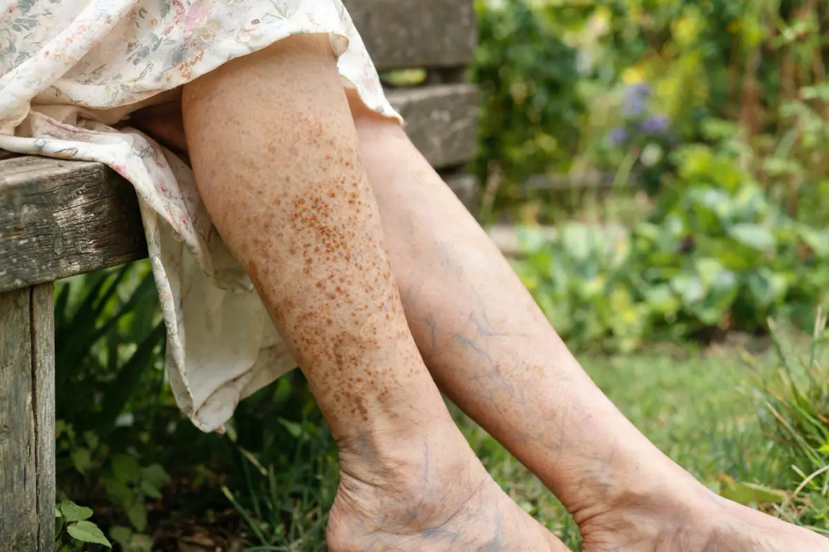
Hemosiderin staining looks like rust-brown, copper or bronze discoloration. On the forearms it appears as brown patches where purple senile purpura used to be. On the lower legs it starts as freckle-like brown dots around the inner ankle and can spread into larger bronze patches, often with swelling or varicose veins. It is flat and doesn't blanch when pressed.
Key takeaways
- Forearm staining follows senile purpura patches [1].
- Leg staining usually starts at the inner ankle and spreads up the shin [2].
- Colour is rust, copper or bronze — not the tan of age spots.
- Hardened, shiny or shrinking skin around stained ankles signals more advanced vein disease [4].
- Any spot that's changing or irregular should be checked for skin cancer.
On this page
The photo at the top shows a typical case on the lower leg. Below, we describe what hemosiderin staining looks like at each stage and in each location, so you can compare with your own skin.
On the forearms and hands
| Stage | What you see |
|---|---|
| Fresh | Dark-purple senile purpura patch, flat, sharp edges [1] |
| Fading (weeks 2–3) | Purple turns duller, brownish at the edges |
| Stain | Rust-brown or tan patch in the same shape as the purple one |
| Long-term | Overlapping brown patches create a bronze or "dirty" tint across the forearm |
The surrounding skin is usually thin, crinkled and sun-damaged, with new purple patches next to old brown ones.
On the lower legs
| Stage | What you see |
|---|---|
| Early | Small brown or copper freckle-like dots around the inner ankle; ankles may swell by evening |
| Established | Dots merge into larger bronze patches spreading up the shin; varicose veins often visible [3] |
| With eczema | Brown skin becomes red, itchy, scaly or weepy [5] |
| Advanced | Skin around the ankle becomes hard, shiny, tight and may shrink like an upside-down champagne bottle (lipodermatosclerosis) [4] |
| Ulcer | Open, slow-healing sore, usually near the inner ankle [2] |
How to tell it apart
| Looks like | Differences |
|---|---|
| Age spots | Tan to dark brown, sharp edges, on sun-exposed skin; not linked to bruising or veins |
| Fading bruise | Yellow-green tones, still tender, disappears in weeks |
| Senile purpura | Purple, not brown — the stage before staining |
| Melanoma or other skin cancer | Irregular border, several colours, growing or changing, may bleed — see a doctor |
| Pigmented purpura (capillaritis) | Cayenne-pepper-like tiny dots, often on legs; a dermatologist can confirm |
What the surrounding skin tells you
The skin around hemosiderin staining gives useful clues:
- Thin, crinkled, sun-damaged forearm skin with purple patches → staining from senile purpura, usually harmless [1]
- Swollen ankles, visible varicose veins → venous staining; worth a vein check [3]
- Itchy, scaly or weeping skin → venous eczema on top of staining [5]
- Hard, tight, shiny skin that feels "woody" → more advanced venous disease [4]
- A shallow, slow-healing wound near the ankle → possible venous ulcer; needs care [2]
Why we describe instead of showing many photos
Skin looks different on every skin tone and in every light, and medical photo libraries mostly show advanced cases. Detailed descriptions help you compare your own skin more fairly. DermNet's image library is a good resource if you want to see more clinical photos.
Taking your own photos
- Same spot, same daylight, once a month
- A coin or ruler for scale
- Both legs, front and inner side
- Note swelling, itch or new sores
Frequently asked questions
What does hemosiderin staining look like?
Rust-brown, copper or bronze discoloration. On forearms, it follows purple patches; on legs, it starts around the ankle and can spread up the shin.
What color is hemosiderin staining?
Usually rust or copper-brown, sometimes bronze or dark brown. It looks different from the tan of age spots.
Is hemosiderin staining raised?
No, it's flat. Raised, hard or lumpy skin with staining suggests more advanced vein disease and should be checked.
Can hemosiderin staining look like a bruise?
It can look like an old, brown bruise that never fully fades. The difference is that it stays for weeks to months and has no tenderness.
Does hemosiderin staining look different on darker skin?
On darker skin tones, staining can look dark brown, grey-brown or almost black rather than rust-coloured, and it may be harder to spot early. The pattern — inner ankle, lower shin, with swelling or varicose veins — is the same.
Can hemosiderin staining appear on the arms?
Yes. On the forearms and hands it follows repeated senile purpura and bruising, appearing as brown patches where purple ones used to be.
Sources
- Senile purpura — DermNet (New Zealand).
- Stasis dermatitis and ulcers — MedlinePlus Medical Encyclopedia.
- Venous insufficiency — MedlinePlus Medical Encyclopedia.
- Caggiati A, et al. Skin iron deposition characterises lipodermatosclerosis and leg ulcer. European Journal of Vascular and Endovascular Surgery, 2010. PubMed 20880725
- Venous eczema — DermNet (New Zealand).
General information, not medical advice. Written by the Reunoa editorial team; Reunoa makes Bruise Defense & Repair Serum. See our editorial policy for how we pick sources and keep product mentions fair.